

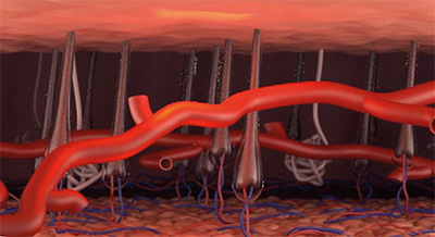
Veins carry blood from the legs back to the heart. The return of venous blood to the heart from the lower limbs is aided with one-way valves in the veins. The contraction of the leg muscles produces a pumping action which squeezes the blood towards the heart during walking or muscular contraction. The valves prevent the blood coming down the leg again.
The venous system in the lower limb is made up of a network of veins consisting of:
Superficial veins:
Are veins located close to the surface of the skin. There are two main trunks called the long saphenous vein and the short saphenous vein as demonstrated in the illustrations. Similar to a tree trunk or a river they have many branches (also called tributaries) which form a network of inter-connecting veins that drain a wide area into the main trunks.
The veins themselves do not have a crucial function and if removed will not result in any problems with the return of the blood to the heart. In fact, these veins are frequently removed in patients with heart disease to use in heart bypass operations.
The two main trunks drain the blood supply of the skin and superficial tissues to the deep veins.
When Vascular Surgeons talk about varicose veins, they refer to these superficial veins which have become dysfunctional and are not working properly.

Deep veins:
Are larger veins located deep in the legs between the muscles and the bones of the legs. These deep veins are the main vessels that return the blood (brought into the legs by arteries) up from the legs to the heart. When talking about deep vein thrombosis (economy class syndrome), these are the ones which are affected.
Perforator (communicating) veins:
Connect the superficial veins to deep veins. They are many in number along the length of the leg but the most important perforators are the sapheno-femoral junction and the sapheno-popliteal junctions at the top of the leg and behind the knee respectively. In the normal physiological setting the direction of flow is from superficial to deep in these veins.
Thread veins/Spider veins/Reticular veins:
These are very small veins (Less than 1.5 mm in diameter) very superficially under the skin which drain into the superficial veins described above. Although part of the normal anatomy, they are not normally visible. They become more frequent with age, and are more common in women especially of the reproductive age and beyond.
Reticular veins tend to be more pronounced than the other two. Strictly speaking, even when apparent they are not referred to as varicose veins; however, it is not unusual to see them in patients with varicose veins as both are fairly common.
Thread veins, Reticular veins, Telangiectasia or Spider veins are termed depending on their size, depth, and configuration. They may become painful or bleed causing bruising of the skin with minimal injury specially in those with blood thinning medications. Most only need treatment for cosmetic reasons. Patients requiring their veins treated may benefit from the CLaCS procedure which combines Laser with injection sclerotherapy using chemical agents.


